
The Ultimate Guide to Understanding the Human Brain in 2024
The brain is a remarkable three-pound organ responsible for regulating all bodily functions, interpreting external information and embodying the essence of our mind and soul. It governs various capabilities including intelligence, creativity, memory, and emotions. Safeguarded within the skull the brain consists of three main parts: the cerebrum, cerebellum, and brainstem.
It processes information received through the five senses- sight, smell, touch, taste and hearing often simultaneously. The brain integrates these sensory inputs to create meaningful perceptions and stores them in memory. It also controls our thoughts, speech, movement, and the functioning of various organs throughout the body.
The central nervous system (CNS) includes the brain and the spinal cord. In contrast, the peripheral nervous system (PNS) comprises the spinal nerves extending from the spinal cord and the cranial nerves emerging from the brain.
Membranes Covering the Brain and Spinal Cord
Meninges cover the whole brain and spinal cord. It has three different layers:
1. Dura Mater
Consist of two layers of dense fibrous tissue. Outer layer lines the skull bones and inner layer covers the brain. These two layers are closely adherent except where the inner layer separates.
- Cerebral Hemisphere- The falx cerebri
- Cerebellar Hemisphere- The falx cerebelli
- Cerebrum and cerebellum- Tentorium Cerebelli
2. Arachnoid Mater
It’s a serous membrane between dura and pia mater- the space b/w dura and arachnoid mater called as subdural space, in which cerebrospinal fluid flows. Arachnoid mater continues to envelope spinal cord and ends by merging the dura mater at the level of 2nd sacral vertebrae.
3. Pia Mater
This is a vascular membrane. Brain covering the convolutions and deepening down into each fissure. This filum terminal pierces the arachnoid and dural tubes and goes on the fuse with the periosteum of coccyx.
Skull
Depending on their shapes, bones are classified as long, short, flat or irregular. Bones are different proportions of the two types of osseous tissue: compact and spongy bone.
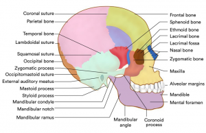

While the former has a smooth structure, the latter is composed of small needle-like or flat pieces of bone called trabeculae, which form a network filled with red or yellow bone marrow. Most skull bones are flat and consist of two parallel compact bone surfaces with layer of spongy bone sandwiched in between. The spongy bone layer of flat bones predominantly contains red bone marrow and has a high concentration of blood.
The skull is a highly complex structure consisting of 22 bones. These can be divided into two sets, the cranial bones and the facial bones. While the latter form the framework of the face, the cranial bones form the cranial cavity that encloses and protects the brain. All bones of the adult skulls are firmly connected by sutures. The frontal bone forms the forehead and contains the frontal sinuses which are air filled cells within the bone. Most superior and lateral aspects of the skull are formed by the parietal bones while the occipital bone forms the posterior aspects.
The base of the occipital bone contains the foramen magnum, which is a large hole allowing the inferior part of the brain to connect to the spinal cord. The remaining bones of the cranium are the temporal, sphenoid and ethmoid bones.
Meninges
The meninges are three connective tissue membranes enclosing the brain and the spinal cord. Their functions are to protect the CNS and blood vessels, enclose the venous sinuses, retain the cerebrospinal fluid and form partitions within the skull. The outermost meninx is the dura mater which enclose the arachnoid mater and the innermost pia mater.
Brain Ventricles and Cerebrospinal Fluid (CSF)
Ventricles: Hollow, fluid-filled cavities in the brain
- Lateral Ventricles: Two ventricles deep within the cerebral hemispheres.
- Third Ventricle: Connects with the lateral ventricles via foramen of Monro.
- Fourth Ventricle: Connects to the third ventricle through the aqueduct of Sylvius.
Choroid Plexus: Ribbon-like structure inside the ventricles that produces clear, colorless CSF.
CSF Flow Path:
- Produced in the choroid plexus.
- Flows from the lateral ventricles to the third ventricle through the foramen of Monro.
- Moves from the third ventricle to the fourth ventricle via the aqueduct of Sylvius.
- From the fourth ventricle CSF flows into the subarachnoid space around the brain and spinal cord.
CSF Reabsorption: Recycled/absorbed by arachnoid villi in the superior sagittal sinus.
Function of CSF: Cushions and protects the brain and spinal cord. Balance constant production and absorption of CSF maintain equilibrium.
Potential Issues: Hydrocephalus enlargement of ventricles due to CSF buildup. Syringomyelia is a collection of fluid in the spinal cord due to CSF flow disruption.
Major Parts of the Human Brain and Their Functions
Major parts of the human brain are the cerebral hemispheres, diencephalon, brainstem and cerebellum.
| Human Brain Regions | Brain Functions |
| Cerebral Hemisphere |
|
| Diencephalon |
|
| Brainstem |
|
| Cerebellum |
|
| Corpus Callosum |
|
| Frontal Lobe |
|
| Parietal Lobe |
|
| Occipital Lobe |
|
| Temporal Lobe |
|
| Limbic System |
|
| Basal Ganglia |
|
Cranial Nerves: Origin, Distribution and Functions
| S. No. | Cranial Nerves | Central Origin | Distribution | Function |
| 1 | Olfactory (sensory) | Smell area in temporal lobe of cerebrum through olfactory bulb | Mucous membrane is roof of nose | Sense of smell |
| 2 | Optic (sensory) | Sight area in occipital lobe of cerebrum, cerebellum | Retina of the eye | Sense of sight balance |
| 3 | Oculomotor (motor) | Nerve cells near the floor of the aqueduct of the midbrain | Superior, interior and medial rectus muscles, and circular muscle, and circular muscle fibers of the iris | Moving the eyeball, regulating the size of the pupils and focusing |
| 4 | Trochlear (motor) | Nerve cells near floor of aqueduct of midbrain | Superior oblique muscles of the eyes | Movement of the eyeball |
| 5 | Trigeminal (mixed) | Motor fibers from the pons varolii sensory fibers from the trigeminal ganglion | Muscles of mastication sensory to gums, cheeks, lower jaw, iris, cornea | Chewing sensation from the face |
| 6 | Abducens (motor) | Floor of fourth ventricle | Lateral rectus muscles of the eye | Movement of the eye |
| 7 | Facial (mixed) | Pons varolii | Sensory fibers to the tongue
Motor fibers to the muscles of the face |
Sensation of tase
Movements of facial expression |
| 8 | Auditory (sensory) | Hearing area of cerebrum | Organ of Corti in the cochlea | Sense of hearing |
| 9 | Glossopharyngeal | Medulla oblongata | Back of tongue and pharynx
Posterior third of tongue Parotid glands |
Sense of tase
Secretion of saliva Movements of pharynx |
| 10 | Vagus (mixed) | Medulla oblongata | Pharynx, larynx, lungs, heart, gallbladder, stomach, small and large intestine | Movement of secretion |
| 11 | Accessory (motor) | Medulla oblongata | Sternomastoid, trapezius, laryngeal, and pharyngeal muscles | Movement of the head and shoulders and pharynx and larynx |
| 12 | Hypoglossal (motor) | Medulla oblongata | Tongue | Movement of tongue |
Blood Supply to the Brain
- Major arteries are vertebral and internal carotid arteries.
- Internal carotid arteries supply to the cerebrum and vertebral arteries supply the cerebellum, brainstem and underside of the cerebrum.
- The two posterior and single anterior communicating arteries form the circle of Willis, equalizes blood pressures in the brain’s anterior and posterior regions and protects the brain from damage, should one of the arteries become occluded.
- There are little communication b/w smaller arteries on the brain’s surface, hence the occlusion of these arteries usually results in localized tissue damage.
Cell of the Brain
Brain is made up of two types of cells: Glia and nerve cells (neurons)
Nerve Cells (Neurons)
1. Structure
- Cell Body (Soma): Contains the nucleus and other organelles; integrates incoming signals.
- Dendrites: Branch-like structures that receive messages from other neurons.
- Axon: Long, slender projection that transmits electrical impulses away from the cell body to other neurons or muscles.
2. Function
- Signal Transmission: Neurons convey information through electrical and chemical signals.
- Electrical Signals: Travel along the axon as action potentials.
- Chemical Signals: Transmitted across synapses using neurotransmitters.
3. Synapse
- Definition: Tiny gap between neurons where communication occurs.
- Transmission:
- Pre-Synaptic Neuron: Sends the signal. Neurotransmitters are released from sacs (vesicles) at the axon terminal.
- Post-Synaptic Neuron: Receives the signal. Neurotransmitters cross the synapse and bind to receptors on the dendrites of the next neuron.
4. Process
- Dendrite Reception: Dendrites pick up chemical messages from other neurons.
- Integration: The cell body processes these messages and decides if they should be passed on.
- Action Potential: If the message is significant, an electrical impulse travels down the axon.
- Neurotransmitter Release: At the axon terminal, neurotransmitters are released into the synapse.
- Signal Reception: Neurotransmitters cross the synapse and bind to receptors on the receiving neuron, continuing the message.
5. Analogy
- Electrical Wiring: Like electrical wiring in a home, where wires carry electricity to power a light bulb, neurons carry electrical impulses to transmit signals.
Glial Cell
Glia cells, derived from the Greek word meaning “glue,” provide essential support to neurons in the brain. They outnumber neurons by a factor of 10 to 50 and play crucial roles in maintaining brain health and function. They are also commonly involved in brain tumors.
1. Astrocytes (Astroglia)
- Regulate the blood-brain barrier, allowing the selective exchange of nutrients and molecules with neurons.
- Maintain homeostasis by balancing ions and neurotransmitters in the brain environment.
- Assist in neuronal defense and repair processes.
- Involved in scar formation following brain injury.
- Influence electrical impulses and synaptic activity.
Structure: Star-shaped with numerous branching processes that interact with neurons and blood vessels.
2. Oligodendrocytes (Oligodendroglia)
- Produce myelin, a fatty substance that insulates axons.
- Myelin sheath increases the speed of electrical message transmission along axons.
Structure: Small cells with fewer processes compared to astrocytes; each oligodendrocyte can myelinate multiple axons.
3. Ependymal Cells
- Line the ventricles of the brain and the central canal of the spinal cord.
- Secrete cerebrospinal fluid (CSF) and help circulate it through the ventricles and around the brain and spinal cord.
Structure: Ciliated cells forming a thin layer that lines the ventricular system.
4. Microglia
- Act as the brain’s immune cells, defending against pathogens and removing debris from damaged or dead cells.
- Engage in synaptic pruning, which helps refine neural connections by eliminating excess or unused synapses.
Structure: Small cells with multiple branching processes that can move through the brain tissue to perform their functions.
Frequently Asked Questions (FAQs)
Q1. Where is brain located?
Ans. The brain is housed inside the bony covering called the cranium. The cranium protects the brain from injury. Together, the cranium and bones that protect the face are called the skull. Between the skull and brain is the meninges, which consist of three layers of tissue that cover and protect the brain and spinal cord.
Q2. Which part of the brain controls memory?
Ans. The functions of memory are carried out by the hippocampus and other related structures in the temporal lobe.
Location: Located within the temporal lobe, part of the limbic system.
Function: Critical for the formation of new memories and spatial navigation. Essential for converting short-term memories into long-term memories.
Q3. What are the functions of the left and right brain?
Ans. The left brain is more verbal, analytical, and orderly than the right brain. It’s sometimes called the digital brain because it’s better at things like reading, writing, and computations. On the other hand, the right brain is more visual, intuitive, and creative.
Related post


