Empowering You
to Become a TopDoc
Our aim is to provide you with the tools and expert support to excel in NEET PG exam and become a top doctor.
Our new app is here! Explore seamless access to courses and features designed to enhance your learning experience. Download Now!


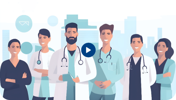
Our faculty consists of experienced professors and specialists with expertise in medical education and practice. Our dedicated faculty provides practical, high-quality lectures to help ensure your success in your medical career.
Know More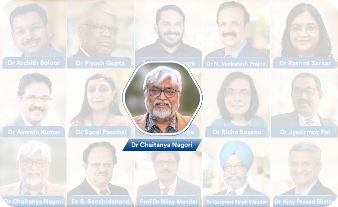
We provide high-quality and up to date medical content created by leading experts. Our resources designed to offer in-depth knowledge, ensuring you to stay ahead in your studies and exam.
Know More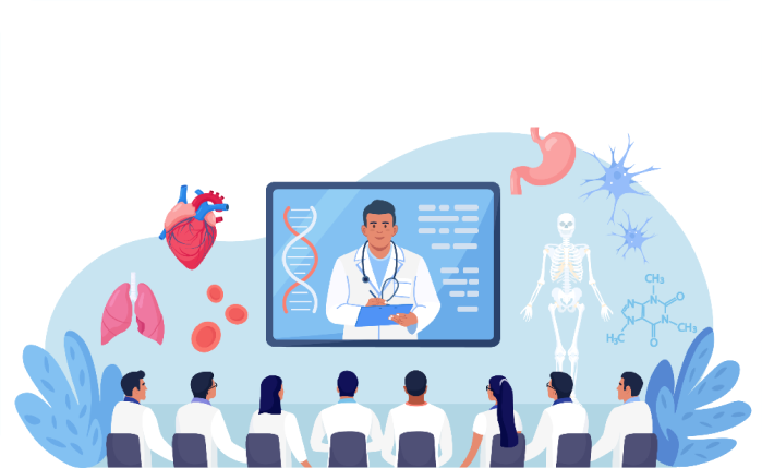
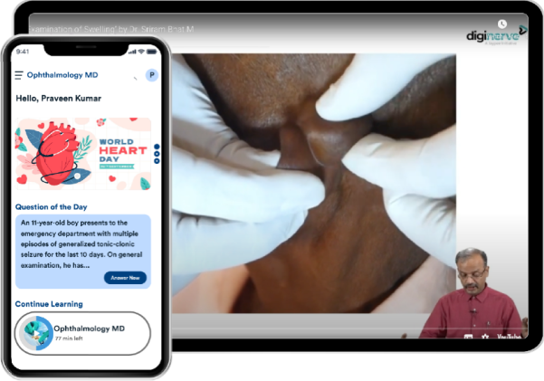
Our courses are specifically tailored for NEET PG and NEET SS preparation featuring expert-curated content, test series, grand tests, QBank, and previous years’ questions (PYQs) designed to help medical students prepare more effectively.
Know More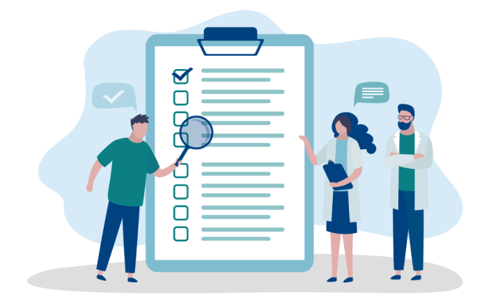
Dr. Wise is the best AI-powered study companion for students in the medical industry. Available at your service 24/7 providing instant answers, offers personalized guidance and clear doubts ensuring students get incomparable support in NEET PG preparation, anytime, anywhere.
Know More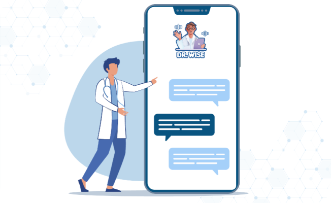
DigiNerve offers exclusive live webinars and chat shows with top medical professionals where you can learn, interact, ask questions and gain insights on key topics. Stay informed with expert guidance to enhance your NEET PG and NEET SS preparation.
Know More

to Become a TopDoc
Our aim is to provide you with the tools and expert support to excel in NEET PG exam and become a top doctor.


“Interactive Learning & Self-Assessment. I love how interactive this e-lecture series is. The self-assessment questions really helped me test my knowledge, and the drug formulary was super useful during my ward rounds. It's a great way to stay updated and reinforce what I've learned.”

“In-Depth Case Discussions. The case discussions in this e-lecture series were incredibly helpful. They made complex dermatology topics so much easier to understand, especially the rare cases I often struggled with. The spotter section helped me a lot in recognizing key lesions for exams.”

“Interactive Learning. The Chat Shows are what make this course unique. It feels like being part of a live classroom with some of the best minds in Pediatrics. These sessions help me stay connected to recent updates and research without feeling overwhelmed.”

“Innovative Features. Dr. Wise, the AI chatbot, is like having a personal tutor 24/7. Whether I'm stuck on a theoretical concept or a clinical scenario, it always provides clear and well-referenced explanations. It's incredibly helpful during my night shifts!”

“Great for Everyday Practice!. I loved how practical this course is. Dr. Piyush Gupta makes everything easy to understand, and I learned so many useful tips for managing pediatric patients. It's perfect for anyone who wants to improve their skills.”

“Focused on Exams and Practical Skills. The Ophthalmology course is exactly what I needed for my exams. It focuses on clinical skills and decision-making, which is so important. The videos and the DxTx tool are great for quick reviews. I feel more confident in both my exams and in treating patients after going through the course.”
With DigiNerve, you can access high-quality medical content at any time, even when you are offline or without an internet connection. Whether at home or on the go, our resources are always ready to support your learning and exam preparation.