


Radial Nerve Injury: Diagnosis and Treatment
Radial nerve is among the first sizeable nerves distributed within the superior limb of the human body. Now let just go through the anatomy of radial nerve before getting into radial nerve injury.
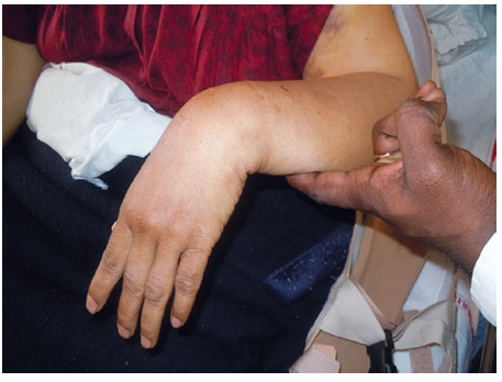
Fig 1: Wrist Drop due to Radial Nerve Palsy
Anatomy of the Radial Nerve
The radial nerve is an important nerve in the arm that mainly controls muscle movement and sensation. Here’s a simpler breakdown of its anatomy:
1. Origin
- The radial nerve starts from the posterior cord of the brachial plexus, which consists of fibers from the cervical spinal nerves C5, C6, C7, C8, and sometimes T1.
2. Pathway
- It travels downward, passing behind the axillary artery.
- It enters the upper arm near the long and medial heads of the triceps muscle, accompanied by the profunda brachii artery.
3. Location in the Arm
- Unlike the spiral groove of the humerus the radial nerve runs alongside the superior aspect of the medial triceps with only muscle fibers separating the nerve from the bone and this is at a distance of 3-4 mm.
- The nerve only encounters the humerus in the lower segment of the bone in a region that passes through the lateral intermuscular septum to the anterior compartment. tissue that keeps it about 3-4 mm away from the bone.
- The nerve only touches the humerus in the lower arm, where it pierces the lateral intermuscular septum to enter the anterior compartment.
4. Branches
- In the upper arm, it gives off motor branches to the triceps.
- About 10 cm above the lateral epicondyle, it enters the anterior compartment between the brachialis and brachioradialis muscles, providing motor branches to the brachioradialis and extensor carpi radialis longus. It also sends a branch to the brachialis, though this muscle is mainly supplied by the musculocutaneous nerve.
5. Bifurcation
- The radial nerve divides into two branches—superficial and deep—around the lateral epicondyle. This can happen anywhere from 4.5 cm above to 4 cm below the epicondyle.
- At this point, it also gives off a branch to the extensor carpi radialis brevis.
6. Superficial Branch
- The superficial branch continues down the arm, underneath the brachioradialis muscle.
- It emerges at the junction of the middle and distal thirds of the forearm, supplying sensation to the skin on the lateral side of the back of the wrist and hand.
7. Deep Branch (Posterior Interosseous Nerve)
- This branch is mainly motor and supplies muscles in the posterior forearm. It passes through the supinator muscle, using a fibrous arch called the arcade of Frohse.
- After exiting the supinator, it quickly divides into branches that innervate:
- Superficial group: Extensor digitorum, extensor digiti minimi, and extensor carpi ulnaris.
- Deep group: Abductor pollicis longus, extensor pollicis longus, extensor pollicis brevis, and extensor indicis.
Injury to the Radial Nerve
It is broadly categorized into four groups: Depending on site where the damage has occurred, which components of the nerve have been affected.
1. Axilla: Radial nerve may be injured in the axilla due to fracture of the proximal humerus and shoulder joint dislocation. Additionally, excessive pressure on the nerve in the axilla, (e.g. poorly fitting crutch can also cause injury).
Motor Functions:
- Triceps brachii and posterior compartment muscles are affected.
- Unable extend at the forearm, wrist and fingers which results in unopposed wrist flexion, referred as wrist drop.
Sensory Functions:
- Four cutaneous branches are affected of the radial nerve.
- Loss of sensation over lateral & posterior forearm.
- Loss of sensation over posterior forearm & dorsal surface of lateral three and a half digits.
2. Radial Groove: Radial nerve is tightly bound within spiral groove of the humerus, it particularly vulnerable to injury in cases of humeral shaft fractures.
Motor Functions:
- Triceps brachii weakened but not paralysed.
- Posterior forearm muscles are affected. Unable to extend at wrist and fingers resulting in unopposed flexion of wrist occurs, known as wrist drop.
Sensory Functions:
- Cutaneous branches to the arm & forearm already formed
- Sensory loss to the dorsal surface to lateral three and half digits, if superficial branch of the radial nerve will be damaged.
- Sensory loss to the associated area on the dorsum of the hand.
3. Forearm: Two terminal branches located within the forearm. Mechanism of injury and effect of their injury differs:
Superficial Branch v/s Deep Branch
Mechanism:
- Superficial Branch: Stabbing or laceration of the forearm.
- Deep Branch: Fracture of the radial head or posterior dislocation of radius.
Motor functions:
- Superficial Branch: None
- Deep Branch: Majority of muscles affected in posterior forearm. Some extension at the wrist maintained and wrist-drop does not occur, due to unaffected extensor carpi radialis longus.
Sensory Functions:
- Superficial Branch: Sensory loss affected the lateral three and half digits and the corresponding area on the dorsum of the hand.
Diagnosis of Radial Nerve Injury
1. Diagnostic Tests for Compression Neuropathies:
- Various tests are available, each with strengths and weaknesses.
- Diagnosis can be achieved through imaging and non-imaging techniques.
2. Non-Imaging Techniques:
Electrodiagnostic Studies:
- Includes electromyography (EMG) and nerve conduction studies (NCS).
- EMG is limited in scope but helpful in ruling out conditions mimicking radial neuropathies.
- NCS is effective in identifying demyelination at the spiral groove.
- Additional techniques may be needed for higher-resolution lesion identification.
- EMG and NCS can diagnose and grade peripheral nerve injuries but may not pinpoint anatomical causes of chronic injuries.
3. Imaging Techniques:
Ultrasonography (US):
- Has improved in resolution over time.
- Limited by operator dependency, leading to variable results.
- High-frequency transducers enhance US effectiveness for specific radial neuropathies, like at the spiral groove.
Magnetic Resonance Imaging (MRI):
- Provides higher soft tissue contrast and reduces user variability.
- High-resolution MR imaging allows differentiation between normal and pathological nerves.
Treatment for Radial Nerve Injury
Medical Management
- Conservative Management:
- First-line treatment for radial nerve entrapment syndrome.
- Includes:
- Oral anti-inflammatory medications.
- Activity modification to avoid aggravating activities.
- Physical therapy, Immobilization with Splinting.
- Limited evidence supporting the effectiveness of these therapies.
- Many studies confirm the need for more effective treatments beyond conservative management.
- Recommended trial of medical management for a minimum of six weeks before progressing to other treatments.
Minimally Invasive Techniques
- Lidocaine Infusion:
- Some discussions around its use for refractory chronic pain.
- Further research needed, particularly regarding piriformis syndrome.
- Corticosteroid Injections:
- Commonly among the first methods attempted.
- Mentioned by multiple authors, but no large-scale studies confirm their efficacy for radial nerve entrapment.
- Peripheral Nerve Stimulation (PNS):
- A newer and effective treatment option.
- Involves implantation of a device under ultrasound guidance.
- Studies indicate significant pain relief in most patients after implantation, though infection risk exists.
- Radiofrequency Techniques:
- Recent advancements noted.
- Pulsed radiofrequency described for treating refractory lateral epicondylitis, showing potential but requiring larger trials for validation.
Surgical Techniques
- General Surgical Interventions:
- Evolving towards smaller incisions and shorter recovery times.
- Recommended only after a trial of 3-6 months of non-operative options.
- Success rates reported up to 92-95% for nerve release procedures.
- Open Surgical Techniques:
- Larger surgical field reduces risk of iatrogenic injuries but involves incisions and scarring.
- Potential complications and opioid use disorder risks noted.
- Endoscopic Techniques:
- Less invasive, reduce post-operative recovery time.
- Comparable efficacy to open techniques regarding pain syndrome, recovery beyond 6 months, and symptom reduction.
- Non-Contact Laser Doppler Flowmetry:
- Utilized during surgery to monitor nerve perfusion in real-time.
- Can reduce incision length and improve functional recovery when used with endoscopic releases.
Frequently Asked Questions (FAQs)
Q1. What happens if the radial nerve is damaged?
Ans. Weakness, loss of coordination of the fingers. Problem straightening the arm at the elbow. Problem bending the hand back at the wrist or holding the hand. Pain, numbness, decreased sensation, tingling, or burning sensation in the areas controlled by the nerve.
Q2. What are the sites of radial nerve injury?
Ans. The radial nerve is prone to entrapment at three different sites: as it passes between the heads of the triceps brachii muscle, in the spiral groove of the humerus, and while piercing the lateral intermuscular septum.
Q3. What is the difference between wrist drop and finger drop?
Ans. The wrist drop is due to the weakness of the extensor carpi radialis longus (ECRL) while the finger and thumb drop due to the fascicular involvement of the posterior interosseus nerve (PIN). Sensory symptoms over the superficial radial sensory along the radial side of the forearm and thumb may also occur.