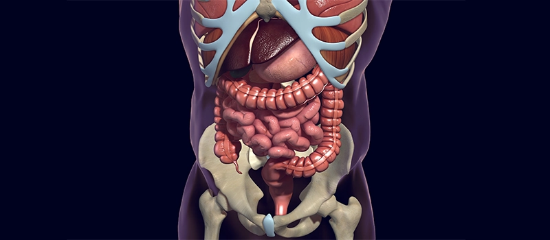
A Comprehensive Guide to Understanding Human Digestive Anatomy
Digestion is defined as the process by which food is broken down into simple chemical substances that can be absorbed and used as nutrients by the body. All the living organisms require nutritive substances and water for survival and growth.
Most of the substances in the diet cannot be utilized as such. These substances must be broken into smaller particles. Then only these substances can be absorbed into blood and distributed to various parts of the body for utilization.
The digestion process is accomplished by mechanical and enzymatic breakdown of foods into smaller chemical compounds. A normal young healthy adult consumes about one kg of solid diet and about 1-2 liters of liquid diet every day. All these food materials are subjected to digestive process before being absorbed into blood and distributed to the tissues of the body.
The digestive system plays major role in the digestion and absorption of the system plays the major role in the digestion and absorption of food substances. Thus, the function of digestive system include:
- Ingestion or consumption of food substances
- Breaking them into small particles
- Transport of the small particles to different areas of the digestive tract
- Secretion of necessary enzymes and other substances for digestion
- Digestion of the food particles
- Absorption of the digestive products
- Removal of unwanted substances from the body
Functional Anatomy of the Digestive System
Digestive System is made up of gastrointestinal tract or alimentary canal and accessory organs, which help in the process of digestion and absorption.
GI tract is a tubular structure extending from the mouth up to anus with a length of about 30 feet. It opens to the external environment on both ends. GI tract is formed by two types of organs:
1. Primary Digestive Organs: This is where the actual digestion takes place. Primary Digestive organs are:
- Mouth
- Pharynx
- Esophagus
- Stomach
- Small intestine
- Large intestine
2. Accessory Digestive Organs: It helps primary digestive organs in the process of digestion. The accessory digestive organs are:
- Teeth
- Tongue
- Salivary glands
- Exocrine part of pancreas
- Liver
- Gallbladder
Tongue
- Functions: Taste, speech, mastication, deglutition
- External Features: Root, tip, vertical surface, dorsal surface, 2 lateral borders
- Dorsal Surface: Covered by mucous membrane with papillae in anterior 2/3rd and lingual tonsil in posterior 1/3rd.
- Ventral Surface: Smooth
- Muscles: Intrinsic (superior longitudinal, inferior longitudinal, transverse, vertical), extrinsic (genioglossus, hyoglossus, styloglossus, palatoglossus).
- Blood Supply: Lingual artery, venae comitantes, deep lingual vein, lingual vein.
- Lymphatic Drainage: Submental nodes, submandibular nodes, jugulo-omohyoid nodes.
- Nerve Supply: Motor-hyoglossal except palatoglossus by pharyngeal plexus sensory-general (lingual-anterior 2/3rd , glossopharyngeal-posterior 1/3rd ), special (chorda tympani-anterior 2/3rd, glossopharyngeal posterior 1/3rd ), posterior most part-vagus.
Salivary Glands
| Features | Parotid Gland | Submandibular Gland | Sublingual Gland |
| Situation | Near external acoustic meatus | Diagastric triangle | Floor of mouth |
| Capsule | Investing layer of deep cervical fascia | Connective tissue | Connective tissue |
| External Features | Apex, surfaces (Base or superior, superficial, anteromedial), borders (anterior, posterior, medial) | Superficial, deep parts | — |
| Structures in Gland | Arteries (External carotid, maxillary superficial temporal), veins (Maxillary superficial temporal, retromandibular), facial nerve with its branches | — | — |
| Duct | Opens into vestibule of mouth opposite upper second molar tooth | Opens into floor of mouth | Opens into floor of mouth |
| Blood Supply | External carotid artery, jugular vein | Facial and lingual arteries, facial and lingual veins | Facial and lingual arteries, facial and lingual veins |
| Nerve Supply | — | — | — |
| Parasympathetic | Glossopharyngeal – otic ganglion- auriculotemporal | Facial- chorda-tympani- submandibular ganglion-lingual | Facial- chorda-tympani- submandibular ganglion-lingual |
| Sympathetic | Plexus around external carotid artery | Plexus around facial artery | Plexus around facial artery |
| Sensory | Auriculotemporal nerve | Lingual nerve | Lingual nerve |
Different Parts of Digestive System
| Organs | External Features | Blood Supply | Nerve Supply |
| Pharynx | Parts (Nasopharynx, oropharynx, laryngopharynx), muscles (Constrictors (superior, middle, inferior), longitudinal (Stylopharyngeus, palatopharyngeus, salpingopharyngeus) | External carotid and maxillary arteries, facial and internal jugular veins | Motor (Pharyngeal plexus except stylopharyngeus by glossopharyngeal nerve), sensory (Glossopharyngeal nerve) |
| Esophagus | 25cm long, extends from cricoid cartilage to stomach, constrictions (From incisor teeth 6 inched, 9 inches, 11 inches, 15 inches) | Arteries (Inferior thyroid, esophageal branches, left gastric) Veins (Brachiocephalic, azygos, left gastric) |
Parasympathetic (Recurrent laryngeal, esophageal), sympathetic (Middle cervical ganglion, upper 4 thoracic ganglia) |
| Stomach | Situation: Epigastric, umbilical, hypochondriac regions. Two orifices: Cardiac, pyloric Two curvatures: lesser, greater Two surfaces: Anterior (liver, diaphragm, left kidney, left suprarenal, pancreas, transverse mesocolon, splenic flexure of colon, splenic artery) |
Arteries: Left gastric, right gastric, left gastroepiploic, right gastroepiploic, short gastric Veins: Superior mesenteric, portal, splenic Lymphatic: Pancreatic splenic, left gastric, right gastroepiploic, splenic, hepatic, pyloric nodes |
Sympathetic: T6-T10 spinal segments Parasympathetic: Vagus |
| Duodenum | Shortest small intestine. 10 inches long. Extent: Pylorus to duodenojejunal flexure 4 parts: first/superior, second/ vertical/descending, third/ horizontal, fourth/ ascending | Arteries: Superior and inferior pancreaticoduodenal Veins: Splenic, superior mesenteric, portal Lymphatics: Pancreaticoduodenal |
Sympathetic: T9, T10 spinal segments Parasympathetic: Vagus |
| Jejunum | Forms upper 2/5th of small intestine. Suspended from fold of mesentery, 1-2 arterial arcades, longer and few vasa recta Extent: Distal jejunum to ileocecal junction. Thick and more vascular walls. | Artery: Superior mesenteric Vein: Superior mesenteric Lymphatics: Superior mesenteric nodes |
Sympathetic: T9, T10 spinal segments Parasympathetic: Vagus |
| Ileum | Forms upper 3/5th of small intestine. Suspended from fold of mesentery, 3-6 arterial arcades, short and more vasa recta Extent: Distal jejunum to ileocecal junction. Thick and more vascular walls | Artery: Superior mesenteric Vein: Superior mesenteric Lymphatics: Superior mesenteric nodes |
Sympathetic: T9, T10 spinal segments Parasympathetic: Vagus |
| Large Intestine | Extent: Ileocecal junction to anus. 1.5 cm long Parts: Cecum and appendix, ascending colon, transverse colon, descending colon, sigmoid colon, rectum, anal canal Taenia coli: thickened longitudinal muscle coats Haustrations/ sacculations: Out pocketing of wall Appendices epiploicae: Appendices epiploicae: Small bags of fats |
Arteries: Superior mesenteric Vein: Superior mesenteric Lymphatics: Superior mesenteric nodes |
Sympathetic: T11- L1 spinal segments upto right 2/3rd of transverse colon rest by L1, L2 spinal segments Parasympathetic: Vagus upto right 2/3rd rest by pelvic splanchnic |
| Cecum | Blind pouch in right iliac fossa. Types: Conical (appendix at apex), funicular (appendix in depression at the center), (appendix in depression at the center), ampullary (appendix at one side) | Arteries: Cecal branches of ileocolic Veins: superior mesenteric |
Sympathetic: T11-L1 spinal segments Parasympathetic: Vagus |
| Appendix | Arises in the posteromedial wall of cecum. 9 cm long Position: Paracolic/11 O’clock, retrocaecal/12 O’clock, splenic/2 O’ clock, sacral/promontoric/3 O’clock, pelvic/ 4 O’clock, midinguinal/6 O’clock |
Artery: Appendicular Veins: Appendicular, ileocolic, superior mesenteric Lymphatic: Ileocolic nodes |
Sympathetic: T9, T10 spinal segments Parasympathetic: Vagus |
| Rectum | Extent: Sigmoid colon to anorectal junction, 12 cm long, Curvatures: 2 anteroposterior, 3 lateral | Artery: superior and middle rectal, median sacral Veins: superior and middle rectal Lymphatic: internal iliac nodes |
Sympathetic: L1, L2 spinal segments Parasympathetic: S2-S4 |
| Anal Canal | 3.8 cm long, 3 parts Upper part: 15mm, mucous membrane thrown into folds (anal columns), attached at lower ends by anal valves (pectinate line), anal sinus is in between anal columns Middle part: 15mm, dense venous plexus Lower part: 8 mm, skin with glands and hair White line of Hilton: Junction between mucous membrane (middle part) and skin (lower part) Sphincters: external, internal |
Artery: Superior and inferior rectal Veins: Internal and External rectal venous plexus Lymphatic: Internal iliac and superficial inguinal nodes |
Sympathetic: L1, L2 spinal segments Parasympathetic: S2-S4 |
| Liver | The largest gland in the body secretes bile, helps in metabolism. Surfaces: Anterior, posterior, right, superior, inferior Lobes: Right, left, caudate, quadrate Porta hepatis: Portal vein, hepatic artery, right and left hepatic duct, hepatic plexus of nerves Ligaments: Falciform, right triangular, left triangular, coronary |
Arteries: Hepatic artery, portal vein Veins: Hepatic sinusoid – interlobar veins – sublobar veins- hepatic veins- inferior vena cava Lymphatics: Hepatic, paracardial, celiac nodes |
Sympathetic: Hepatic plexus Parasympathetic: Vagus |
| Pancreas | Partly exocrine and endocrine gland Extent: Duodenum to spleen Parts: Head, neck, body tail Ducts: Main pancreatic (duct of Wirsung), accessory pancreatic duct opens into major and minor duodenal papilla. |
Arteries: Branches of splenic, superior and inferior Pancreaticoduodenal Veins: Splenic vein Lymphatics: Pancreatic splenic, celiac, superior mesenteric nodes |
Sympathetic: Splanchnic nerves Parasympathetic: Vagus |
Different Parts of Digestive System and its Functions
| Organ | Function |
| Pharynx | The pharynx directs air from your nose and mouth to the larynx (voice box) which then passes air to the trachea and lungs. It also channels food and liquids to the esophagus, which sends them to your stomach. Additionally, the pharynx helps prevent food and liquid from entering your trachea and lungs. |
| Esophagus | The esophagus is a muscular tube that functions to transport food and liquids from the mouth to the stomach. It moves these substances through a series of coordinated muscle contractions called peristalsis. |
| Stomach | Reservoir of food. Softens and mixes the food with the gastric juice. Gastric glands produce gastric juice, HCL. |
| Duodenum | Duodenum is the first part of the small intestine and plays a crucial role in digestion. Its primary functions are:
|
| Jejunum | Jejunum is the middle section of the small intestine, located between the duodenum and the ileum. Its primary functions include:
|
| Ileum | Ileum is the final section of the small intestine, located between the jejunum and the large intestine. Its primary functions include:
|
| Large Intestine | Absorption of water and storage of matter reaching it from the small intestine. Bacteria present in the large intestine helps to synthesize Vitamin B. |
| Cecum | Cecum is the first part of the large intestine, located in the lower right abdomen where the small intestine meets the large intestine. Its primary functions include:
|
| Appendix | Appendix is a small, tube-like structure attached to the cecum of the large intestine. Its function include:
|
| Rectum | Rectum is the final section of the large intestine, leading from the sigmoid colon to the anus. Its primary functions include:
|
| Anal Canal | Anal canal is the final segment of the digestive tract, located between the rectum and the anus. Its primary functions include:
|
| Liver | Liver is a vital organ with wide range of functions, including:
|
| Pancreas | Pancreas is vital organ with both endocrine and exocrine functions:
|
Frequently Asked Question (FAQs)
Q1. What role do smooth muscles play in digestion?
Ans. Smooth muscles in the digestive tract help move food through peristalsis, ensuring it progresses from the esophagus to the stomach and intestines.
Q2. How does the gall bladder contribute to digestion?
Ans. The gall bladder stores and concentrates bile, which is released into the small intestine to aid in the chemical breakdown of fats.
Q3. What happens to undigested food in the digestive system?
Ans. Undigested food moves into the large intestine, where water is absorbed, and the remaining material is eventually expelled as stool during bowel movements.
Q4. How do pancreatic enzymes aid digestion?
Ans. Pancreatic enzymes, such as amylase and lipase, break down carbohydrates and fats in the small intestine, facilitating the absorption of amino acids and other nutrients.
Q5. What is the function of the esophageal sphincter?
Ans. The esophageal sphincter controls the passage of food from the esophagus to the stomach, preventing acid reflux and ensuring proper digestion.
Related post
