A Detailed Overview of Various Ventilator Modes in ICU
Overview
Ventilation is an important area of respiratory care and aims to ensure the proper gas exchange within the lungs.
The respiratory and cardiovascular systems collaborate to supply oxygen O2 to tissues and release carbon dioxide, CO2.
When patients experience respiratory difficulties, there is often an elevation in CO2 levels in their blood gases.
Strategies to address this include airway clearance through suction and mechanically augmented respiratory parameters such as atmospheric pressure rate, respiratory rate, pressure, or volume to enhance ventilation.
To achieve the required pressure for mechanical ventilation, access to the airway is imperative. This access is typically facilitated through methods such as:
- Oral or nasal endotracheal tube (ETT)
- Tracheostomy Tube
- Well-sealed mask for non-invasive ventilation
These methods ensure efficient delivery of ventilatory support, thereby aiding in maintaining adequate gas exchange in the lungs.
There are two primary common modes of ventilation:
- Invasive Ventilation: Utilized for unconsciousness patients.
- Non-invasive Ventilation: Suitable for conscious patients.
These various techniques of mechanical ventilation are important topics for exams like NEET-PG/next and FMGE.
What are the different types of ventilator modes?
Different methods/modes of ventilation, offers tailored treatment plan that respond to the patient’s specific pathology and requirements.
| Controlled Modes | Supported Modes | Combined Modes | Spontaneous Breathing |
| Volume Control | Pressure Support | AUTOMODE: Volume Control- Volume Support | Continuous Positive Airway Pressure |
| Pressure Control | Volume Support | AUTOMODE: Pressure Control – Pressure Support | Nasal Continuous Positive Airway Pressure |
| Pressure-Regulated Volume Control | Non-Invasive Ventilation- Pressure Support | AUTOMODE: Pressure-Regulated Volume Control – Volume Support | |
| Non-Invasive Ventilation – Pressure Control | Synchronized Intermittent Mandatory Ventilation: Volume Control+ Pressure Support | ||
| Synchronized Intermittent Mandatory Ventilation: Pressure Control+ Pressure Support | |||
| SIMV: Pressure-Regulated Volume Control+ Pressure Support |
There are different types of Ventilator modes which are divided up into pressure or volume-controlled modes, this modern approach classifies ventilatory modes based on three characteristics: the triggers (Flow versus pressure), the limit (what determines the size of the breath), and the cycle (What ends the breath).
1. Controlled Mandatory Ventilation (CMV)
Also known as Assist-Control Ventilation, is a mode of mechanical ventilation where each mandatory breath is either as assist or control breath, all delivered with the same preset volume or partial pressure.
This mode is particularly suitable for patients who require minimal breathing effort, as the ventilator fully controls the patient’s total breathing. CMV is indicated in patients with severe neurological alterations, deep sedation, shock, or respiratory failure. CMV is a common way to decrease the intracranial pressure after head injury.
It ensures consistent ventilation regardless of the patient’s inspiratory efforts. It’s crucial to note that CMV does not eliminate work of breathing entirely, as the diaphragm may still be active.
Therefore, patients should be heavily sedated, and drugs like fentanyl, dexmedetomidine, or midazolam can be used to achieve this level of sedation.
Features of Controlled Mandatory Ventilation:
- Physical Character: Pressure is being controlled.
- Tidal Volume: Tidal Volume is around 7-8ml/Kg.
- Patient Efforts: No efforts from the patient’s side.
- Usage: Typically used in heavily sedated patients.
- Effect on BP and Urine Output: May lead to decrease in BP and urine output due to controlled ventilation.
2. Assist-Control Ventilation
It is a mode of mechanical ventilation where each mandatory breath is either an assist or control breath, all delivered with the same preset volume or partial pressure.
This mode is particularly suitable for patients who require minimal breathing effort, as the ventilator fully controls the patient’s total breathing. CMV is indicated in patients with severe neurological alterations, deep sedation, shock, or respiratory failure. CMV is a common way to decrease intracranial pressure after a head injury.
It ensures consistent ventilation regardless of the patient’s inspiratory efforts. It’s crucial to note that CMV does not eliminate the work of breathing, as the diaphragm may still be active.
Therefore, patients should be heavily sedated, and drugs like fentanyl, dexmedetomidine, or midazolam can be used to achieve this level of sedation.
Features of Controlled Mandatory Ventilation:
- Physical Character: Pressure is being controlled.
- Tidal Volume: Tidal Volume is around 7-8ml/Kg.
- Patient Efforts: No efforts from the patient’s side.
- Usage: Typically used in heavily sedated patients.
- Effect on BP and Urine Output: This may lead to a decrease in BP and urine output due to controlled ventilation.
3. Synchronized Intermittent Mandatory Ventilation (SIMV)
SIMV is a ventilator mode that offers partial mechanical assistance while allowing the patient to breathe spontaneously.
Unlike Assis-control ventilation, in SIMV, the patient’s breaths are partially on their own, reducing the risk of hyperinflation or alkalosis. Mandatory breaths are synchronized with spontaneous respirations, providing support when needed.
SIMV may increase the work of breathing and reduce cardiac output, potentially prolonging ventilator dependency. The addition of pressure support to spontaneous breaths can alleviate some of the work of breathing.
SIMV is often used as a weaning mode, allowing patients to gradually regain their respiratory function. Moderate sedation is typically required to ensure patient comfort and synchronization with the ventilator.
SIMV is indicated for conditions such as high risk of hyperventilation/respiratory alkalosis due to increased respiratory rate, pulmonary edema, acute respiratory distress syndrome, neuromuscular disorders, and cardiac thoracic surgery. It’s typically avoided in patients with shock and head injuries.
Features of Controlled Synchronized Intermittent Mandatory Ventilation:
- Physical Character: Respiratory rate (RR) is 14/min.
- Tidal Volume: Tidal Volume is 400ml/Kg.
- Patient Efforts: Allows for some spontaneous sedation.
- Usage: Can be used in patients with no or slight sedation.
- Effect on BP and Urine Output: May have less impact on BP and urine output due to less protective ventilation compared to CMV.
4. Pressure Control Ventilation
It is a common ventilator mode that offers less risk of barotrauma compared to assist control ventilation and SIMV, as it does not allow for patient-initiated breaths. In PCV, the respiratory flow pattern decreases exponentially, reducing peak pressures and improving gas exchange.
However, there are no guarantees for volume especially when lung mechanics are changing, making it traditionally preferred for patients with neuromuscular disease but otherwise normal lungs.
Pressure is fixed manually, and the ventilator decides the volume. PCV is mainly used for conditions like Acute Respiratory Distress Syndrome (ARDS). A major drawback of this mode is the risk of endotracheal tube obstruction due to secretions, leading to reduced volume reaching the patient’s lungs and resulting in respiratory acidosis due to poor oxygenation and increased CO2 levels.
Features of Pressure Control Ventilation:
- Physical Character: Operates on pressure control, maintaining set inspiratory pressure.
- Tidal Volume: Not designed specifically as a weaning mode.
- Patient Efforts: Facilitates spontaneous breathing efforts.
- Usage: Commonly employed in pediatric patients due to its adaptability.
- Effect on BP and Urine Output: May exert less influence on blood pressure and urine output as it allows for spontaneous breathing, potentially improving oxygenation without significantly impacting cardiovascular function.
5. Pressure Support Ventilation
PSV is a spontaneous mode of ventilation where each breath is initiated by the patient but supported by constant pressure inflation.
It operates two mechanisms: CPAP (Continuous Positive Airway Pressure) and PEEP (Positive End-Expiratory Pressure), which helps open the alveoli. PSV allows the patient to determine inflation volume and respiratory rate, although pressure remains controlled by the ventilator.
Therefore, it can only augment spontaneous breathing, typically with pressures ranging from 5-10cm H2O, especially during weaning. PSV can be delivered through specialized face masks.
Features or Pressure Support Ventilation:
- Physical Character: Works on pressure control
- Tidal Volume: Not a weaning mode.
- Patient Efforts: Facilitates spontaneous breathing.
- Usage: Often used in pediatric patients.
- Effect on BP and Urine Output: This may have less impact on BP and urine output, as it allows for spontaneous breathing and may improve oxygenation.
6. Volume Control Ventilation
In the mode of ventilation, the ventilator delivers a predetermined tidal volume to the patient with each mandatory breath. This mode is typically synchronized with the patient’s inspiratory effort, ensuring that the desired tidal volume is consistently delivered. Volume control ventilation is commonly used in patients with normal lung compliance and resistance, as it helps maintain a consistent ventilation pattern.
Another mode of volume ventilation is Assist-Control Ventilation (ACV). ACV combines the features of volume control ventilation with the ability to support spontaneous breathing efforts. In this mode, the ventilator delivers a set tidal volume with each breath initiated by the patient.
Features of Volume Control Ventilation:
- Physical Character: Operates on volume control, delivering a predetermined tidal volume.
- Tidal Volume: Delivers a set tidal volume.
- Patient Efforts: Patient effort can initiate the breath, but the ventilator ensures that the desired tidal volume is delivered.
- Usage: Commonly used in patients with normal lung compliance and resistance.
- Effect on BP and Urine Output: Volume control ventilation may increase intrathoracic pressure, which can have an effect on blood pressure and urine output. However, the impact can vary depending on the patient’s condition.
7. PEEP (Positive End-Expiratory Pressure)
PEEP level helps to keep the alveoli open at the end of expiration and helps in increasing partial pressure, thereby aiding in improving patient oxygenation. This increase in intrathoracic pressure typically leads to a reduction in venous pressure and carbon dioxide levels. However, it is essential to note that while a decrease in blood pressure might occur, it doesn’t necessarily always result in decreased urine output. Positive End-Expiratory Pressure, expressed in centimeters of water (cmH2O), applies pressure at the end of exhalation, thereby preventing the air sacs in the lungs from collapsing and further improving oxygenation.
8. CPAP (Continuous Positive Airway Pressure)
It is commonly employed to assess a patient’s readiness for extubating, particularly when minimal ventilation support is required.
It maintains a constant circuit pressure as specified by the operator throughout ventilation. Pressure Support Ventilation is often combined with CPAP, providing positive pressure assistance throughout the breathing cycle.
PSV can be delivered through a mask and is utilized in conditions such as obstructive sleep apnea, especially when utilizing a nasal mask.
Additionally, it can be used to delay intubation or manage acute exacerbations of Chronic Obstructive Pulmonary Disease, respiratory distress syndrome, or acute lung injury.
9. Airway Pressure Release Ventilation
It delivers a constant high artificial airway pressure to ensure oxygenation, while ventilation occurs through the release of that pressure.
During the majority of the cycle, a continuous high pressure is applied for a set duration followed by a brief period of lower pressure. The concept revolves around maintaining constant alveolar volume during the extended T high phase (covering 80%-90% of the cycle), enhancing oxygenation.
This extended period of high pressure, often termed an open lung strategy minimizes the repetitive inflation and deflation of the lungs observed in other ventilation modes, thus mitigating the risk of ventilator-induced lung injuries.
APRV offers a unique approach to optimizing respiratory mechanics in critically ill patients in the intensive care unit, reducing the respiratory effort required for ventilation.
Frequently Asked Questions (FAQs)
Q1. What are potential complications associated with the intermittent Mandatory Ventilation mode?
Ans. One notable complication of IMV is breath stacking, characterized by a spontaneous breath occurring immediately after a mechanical breath. This sequence can elevate Peak Inspiratory Pressure (PIP), posing a risk of barotrauma and cardiac compromise.
Q2. How do ventilators work in the Intensive Care Unit ICU?
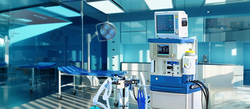
Ans. In the ICU, mechanical ventilators, provide positive pressure ventilation, adjusting ventilator settings like inspiratory time and respiratory cycles to optimize oxygenation. They support patients with respiratory failure while considering factors such as venous return and the effects of positioning, like the prone position, to improve the ventilation-perfusion ratio.
Q3. What happens when the ventilator pressure goes to zero during mechanical ventilation?
Ans. When the ventilator pressure drops to zero, the elastic recoil of the lungs pushes air out. However, the time allotted for exhalation may not be sufficient for all the air to leave the lungs completely.
Q4. What are the advantages of Pressure Control Ventilation?
Ans. Pressure control ventilation offers several advantages, it allows for precise control of alveolar pressure, promoting lung protective ventilation strategies. This ventilatory mode enables adjustment of inspiratory flow rates and initial ventilator settings tailored to individual needs. Respiratory therapists can optimize therapy by titrating the pressure support level, ensuring a favourable pressure gradient for adequate gas exchange.
Related post


