Surgical Techniques for Chronic Sinusitis
Chronic sinusitis is a chronic inflammation of mucous membranes of paranasal sinuses by which irreversible degenerative changes have occurred. Almost invariably succeeds acute sinusitis which did not receive adequate treatment, or it can also develop following a cold or tooth infection.
It occurs when the self-cleansing mechanism of nose and paranasal sinuses gets impaired. Most involved sinusitis is maxillary sinus with duration of symptoms is more than 3 months.
Etiology
Causes of chronic sinusitis are:
- Infection of pharynx, nose and molar teeth
- Trauma to the sinuses and barotraumas
- Local factors include deviated nasal septum, allergy and nasal polypi
- Also includes, chest conditions, such as asthma, chronic bronchiectasis, and chronic bronchitis, responsible for chronic sinusitis.
Chronic sinusitis according to histological changes in the sinus mucosa as follow:
1. Atrophic Sinusitis
Main changes take place in afferent vessels leading to cellular response at and around the arterioles and arteries, later the vessel wall itself becomes thickened and contracted causing endarteritis and thrombosis. In this condition, there is much less edema present as this is primarily a condition that affects the horse’s lower jaw. Hypertrophic and atrophic coexist in the same sinus, the condition causing atrophy at one location and polypoidal hypertrophy at the other place.
2. Hypertrophic Sinusitis
It is characterised mainly by the fact that inflammation is chiefly of the efferent vessels and of the lymphatics. Recurrent stresses take place, which result in changes of the venous and lymphatic flow and organization lead to the formation of oedema and polypoidal mucus membranes, polyps, oedema of periosteum and osteoporosis.
3. Papillary Sinusitis
Occurs when metaplasia of ciliated columnar epithelium to stratified squamous type and throughout the papillary hyperplastic epithelial cells or stroma may be seen inflammatory cells. It is a viral infection.
4. Follicular Sinusitis
Small follicles are seen in the mucous membranes of the sinuses.
5. Glandular Sinusitis
Increase markedly in the submucosal tissue lining of sinuses.
What Kind of Surgery is Done for Chronic Sinusitis?
There are different types of surgery including minimally invasive techniques using endoscopes to remove blockages such as polyps or infected tissue, or to improve drainage in the sinuses. Here are some surgical procedures for chronic sinusitis:
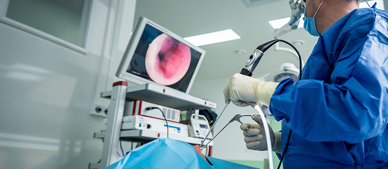
Functional Endoscopic Sinus Surgery
Functional endoscopic surgery is a procedure to re-establish the drainage of the natural ostia and to restore ventilation and mucociliary clearance.
It is based on the principle that clearing the blocked ostium will restore the mucociliary clearance and the diseased mucosa normalizes.
Equipment Used for FESS
- 4 mm 0-degree endoscope
- Angled endoscopes: 30◦, 45◦, 70◦
- Camera
- Display screen
- Light source
Indications for Endoscopic Sinus Surgery
- Chronic Sinusitis
- Nasal Polyps
- Sinus Tumors
- Anatomical Abnormalities
Procedure
- First stack system positioned infront of surgeon. Usually done under general anesthesia, some surgeons prefer local anesthesia especially is unfit patients. Decongestion is done in the observation room with pledgets or nasal patties.
- Patient lies in supine position with head on a ring and head end can be elevated to 15 – 30 degrees.
- The two techniques are:
- Stammberger’s technique (anterior to posterior): Surgery is done from uncinate process towards sphenoid sinus.
- Wigand’s technique (posterior to anterior): Surgery starts from sphenoid sinus and proceeds anteriorly.
- The pledgets/patties soaked in 4% xylocaine adrenaline are removed and a thorough endoscopic examination is done with the three passes.
- First pass, between the septum and inferior turbinate up to choana to visualize the nasopharynx and Eustachian tube.
- In second phase, it passes through middle meatus.
- In third phase, between the superior turbinate and the septum up to the visualization of sphenoid ostia.
- Local infiltration using 2% lignocaine adrenaline given on the axilla of middle turbinate, septum, uncinate process, middle turbinate and lateral wall.
- Uncinate process is identified and the uncinectomy is done.
- Maxillary ostia are identified, widened and the maxillary sinus is cleared.
- Clearance of the anterior ethmoids beginning with the bulla ethmoidalis then done.
- Posterior ethmoids are then cleared after removal of the basal lamella and cleared.
- If there is involvement of the frontal sinus, then the frontal recess is cleared. If there is isolated frontal sinus involvement, it can be accessed without removing the bulls, called as the intact bulla technique.
- Sphenoid sinus can then be approached via the inferomedial aspect of the most posterior ethmoid cell.
- It can also be approached medially by identifying its ostium around 1.5 cm above the roof of the nasopharynx.
- After completion of surgery and achieving hemostasis, nasal packing is done.
Balloon Catheter Sinuplasty (BCS)
Ballon Sinuplasty is a minimally invasive procedure used to treat chronic sinusitis. It includes use o a ballon catheter to dilate the sinus openings, improving drainage and airflow.
Balloon sinuplasty is a medical treatment that is employed by ear, nose, and throat surgeons to open blocked sinus, especially the sinusitis patients who do not respond to drugs.
The United States Food and Drug Administration approved this endoscopic, catheter-based procedure for chronic sinusitis in 2005. It employs the use of a balloon inflated over a wire catheter in order to open up the sinuses passages. It therefore helps to regain normal drainage because when filled the balloon stretches the sinus opening and therefore the walls of the passageway.
Indications:
- Chronic Sinusitis
- Nasal Obstruction
Procedure:
- Patients undergo imaging such as CT scan to assess the sinus anatomy.
- Performed under local anesthesia, sometimes with sedation.
- An endoscope is inserted into nasal passage with small balloon catheter which is threaded into the blocked sinus cavity.
- Balloon is inflated to widen the sinus opening and the balloon is deflated, removed left the passage open.
Frequent Asked Questions (FAQs)
Q1. What are the different types of sinus surgery?
Ans. Here are some different types of sinus surgery:
- Functional endoscopic sinus surgery (FESS)
- Turbinate surgery
- Balloon sinus dilation
- Adenoidectomy
Q2. What is the conservative treatment for chronic sinusitis?
Ans. Chronic sinusitis with polyps should be treated with topical nasal steroids. If severe or unresponsive to therapy after 12 weeks, a short course of oral steroids can be considered. Leukotriene antagonists can be considered.
Q3. What are the differences between Functional Endoscopic Sinus Surgery (FESS) and balloon sinuplasty?
Ans. FESS is a more traditional approach that involves the endoscopic removal of obstructive tissue and polyps to restore sinus drainage. In contrast, balloon sinuplasty is a less invasive technique that utilizes a balloon to dilate the sinus openings without extensive tissue removal. Both techniques aim to improve sinus drainage, but their applications may vary based on the severity and anatomy of the sinus disease.
Q4. What are the potential complications and considerations during the post-operative period for sinus surgery?
Ans. Potential complications include bleeding, infection, and cerebrospinal fluid leaks, although these are relatively rare. Post-operative care involves monitoring for signs of complications, managing pain, and ensuring proper nasal hygiene. Medical students should be aware of the importance of follow-up evaluations to assess healing and address any complications early. Educating patients on signs of complications is also a vital part of post-operative care.
Related post
