Important Topics for Radiology
Radiology is a study of medical technology, it is a important discipline included in the MBBS typically introduced in the later years of medical training. It covers various imaging techniques used to diagnose and manage diseases, including X-ray, ultrasound, computed tomography, positron emission tomography, nuclear medicine and magnetic resonance imaging.
Radiology serves as a critical link between clinical medicine and diagnostic image technology helping medical professionals to visualize internal structures and identify pathological conditions.
A strong grip in radiology for medical students is important, as it aids in accurate diagnosis, treatment/management planning and monitoring of diseases progression with stages.
The radiology curriculum in MBBS covers topics like principles of imaging modalities, anatomy in imaging, interpretation of radiographs, advanced imaging techniques, radiation safety, and the role of radiology in various clinical scenarios, including trauma, oncology, and pediatric care.
Important Topics in Radiology
In the NEET-PG examination 10- 15 questions are asked, while in the INI-CET there are about 15-20 questions that are based on the Radiology Subject.
These competitive exams mainly cover the extent of the candidates’ knowledge about medical imaging procedures, diagnosis diagnostic accuracy principles and role of radiology in clinical practice.
Acquaintance of subject weightage, typical examination formats, areas of significant potential for high yield and general study strategies can greatly help to improve conceptual understanding of Radiology exam.
Imaging of All Emergencies
- Pneumothorax
- Tension Pneumothorax
- Pneumomediastinum
- Pneumoperitoneum
- Pneumocephalus
- Aortic Dissection
- Aortic Aneurysms and Rupture
- Pseudoaneurysms-Yin yang sign
- Pulmonary Thromboembolism Stroke Imaging-Acute Infarct
- Hyperdense MCA sign
- DWI
- Head Trauma-Epidural hematoma
- Swirl sign
- Subdural hematoma
- Subarachnoid hemorrhage
- Intraparenchymal and intraventricular bleed Abdominal Trauma-FAST
- CECT liver lacerations
- Splenic injury Acute Abdomen-Acute Pancreatitis
- Small and large Intestinal obstruction and Volvulus
X-Rays
Concepts of Kilovolt Peak (KVP) and Milliampere-Seconds (MAS)
- KVP (Kilovolt Peak): Refers to the maximum voltage applied across the X-ray tube, influencing the quality and penetrability of the X-ray beam. Higher KVP results in better image quality with less radiation exposure.
- MAS (Milliampere-Seconds): Indicates the quantity of X-ray exposure, combining the current (mA) and the duration (s) of the exposure. It affects the density and contrast of the image.
Important X-ray Views
- Water View
- Caldwell View
- Rhese View
- Stryker’s View
- Schuller View
- Lordotic View
- Reverse Lordotic View
Radiation Interactions
- Compton Effect
- Photoelectric Effect
- Bremsstrahlung Radiation
Mammography Technique
- Differences from Conventional Radiography
Hysterosalpingography Images
- Normal
- Unicornuate Uterus
- Bicornuate Uterus
- Didelphys Uterus
- Hydrosalpinx
IVP Images
- Ureterocele
- Droopy Lily Sign
- Retrocaval Ureter
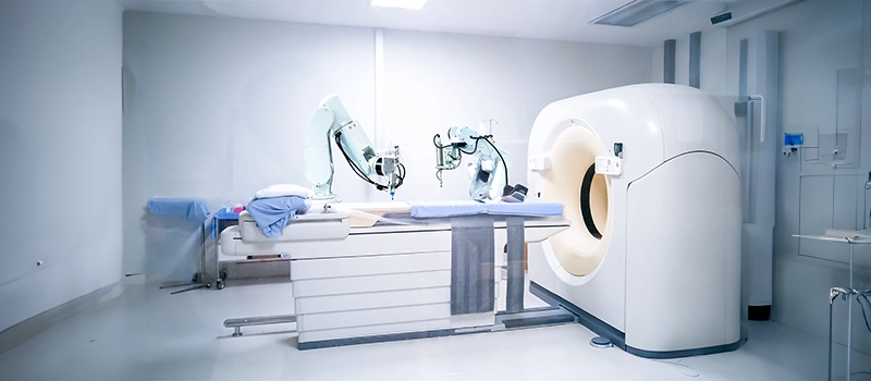
CT Scan
Types of CT Imaging
- Spiral CT
- HRCT (High-Resolution CT)
- MDCT (Multidetector CT)
- Dual Energy CT
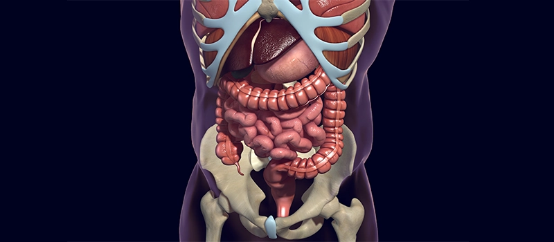
CT Anatomy
- Brain
- Mediastinum
- Abdomen
- Lungs
Coronary Calcium Scoring
- Agatston Scoring
CT Angiography
- Pulmonary Thromboembolism
Radiation Protection
- Lead Apron
- TLD Badge (Thermoluminescent Dosimeter)
MRI Indications and Contraindications
- MRI Sequences: T1-weighted imaging, T2-weighted imaging, FLAIR (Fluid-Attenuated Inversion Recovery), STIR (Short Tau Inversion Recovery), DWI (Diffusion-Weighted Imaging), DTI (Diffusion Tensor Imaging).
- MR Spectroscopy
- MRI Planes: Axial Images, Coronal Images, Sagittal Images of brain
USG
- Piezoelectric Effect: General of electrical charge in certain materials under mechanical stress
- Ultrasound Phenomena: Posterior Acoustic Shadowing, Posterior Acoustic Enhancement
- FAST (Focused Assessment with Sonography for Trauma
- EFAST (Extended FAST)
- EUS (Endoscopic Ultrasound
- Doppler Ultrasound Techniques: Color Doppler, Spectral Doppler
- Doppler Assessments: Umbilical Artery Doppler, Uterine Artery Doppler, Fetal MCA (Middle Cerebral Artery) Doppler
Radiotherapy
1. Teletherapy
- Linac
- Stereotactic Radiotherapy
- IMRT (Intensity-Modulated Radiation Therapy)
- Craniospinal Irradiation
- Electron Beam
- Proton Beam: Bragg Peak
2. Brachytherapy
- Permanent and Temporary Implants
- Pura Beta Emitters
3. Systemic Radiotherapy
- I-131
- Strontium-89
- P-32
4. Law of Bergonie and Tribondeau
5. Radiosensitivity of Tissues and Tumors
6. Different Iodine Isotopes
- I-131
- I-125
- I-124
- I-123
7. Half-Lives of Important Radioisotopes
- F-18
- Tc-99m
- Iodine Isotopes
- P-32
- Co-60
- Cs-137
Nuclear Medicine
- Thyroid Imaging: Thyroid Scintigraphy, Lingual Thyroid
- Renal Scans: DMSA, DTPA, MAG-3 Scan
- Cardiac Imaging: Myocardial Perfusion Imaging, Myocardial Infarct Imaging
- Bone Imaging
- Sulfur Colloid Scan
- Tc-99m Sestamibi Scan
- Octreotide/Somatostatin Receptor Scintigraphy
- PET Imaging
- HMPAO-SPECT
Neuroradiology
- Imaging of Meningioma
- Tumor Comparisons: Medulloblastoma vs. Ependymoma, Arachnoid Cyst vs. Epidermoid Cyst, Craniopharyngioma vs. Pituitary Adenoma
- Important Named Signs: Mount Fuji Sign, Racing Car Sign, PAND Sign, Hummingbird Sign
- CNS Conditions: TB Meningitis, Creutzfeldt-Jakob Disease (CJD)
- Imaging of Stroke: Hyperdense MCA Sign, Penumbra, CT Perfusion Imaging
- Intracranial Bleeds: Extradural Bleed, Subdural Bleed, Subarachnoid Bleed, Intraventricular Bleed, Intraparenchymal Bleed
Respiratory Radiology
- X-ray Views: Posteroanterior vs. Anteroposterior View
- Medical Conditions: Collapse, Consolidation, Pleural Effusion, Pneumothorax
- Important Signs: Golden S Sign, Luftsichel Sign, Silhouette Sign
- Specific Conditions: X-ray of Pulmonary Edema, Sarcoidosis, Pulmonary Thromboembolism
- CT Imaging: Bronchiectasis, Interstitial Lung Disease, Pulmonary Alveolar Proteinosis
- Fungal and Parasitic Infections: Aspergillosis, Hydatid Disease of the Lung, Lung Abscesses, Fungus Ball, Hydropneumothorax
- Other Findings: Lucent Hemithorax, Foreign Body
Frequently Asked Questions (FAQs)
Q1. Is radiologist a good career in India?
Ans. Radiology is one of the most sought-after medical specialities and is in high demand globally. One needs at least 7 years of formal medical education to become a radiologist in India.
Q2. What is the highest paid job in radiology?
Ans. 7 highest-paying radiology jobs are:
- MRI technologist
- Radiologic technologist
- Cardiovascular technologist
- Sonographer
- Radiation therapist
- Nuclear medicine technologist
- Ultrasonographer
Q3. What is the salary of a Radiologist in India?
Ans. Radiologists make an average salary of INR 4,00,000 per year. Entry-level job positions offer around INR 3,00,000. The experienced workers make up to INR 2,975,000 per year.